


Chondral Injury
Damage to the joint surfaces
The knee joint is the largest synovial joint in the body. The ends of the bones are covered in hyaline cartilage and form the articular surfaces.
Hyaline cartilage has very low friction and is lubricated by synovial fluid (from the joint lining)
The joint surfaces can be injured and an area of cartilage can be lost
Osteochondral injury is an injury to the joint surface (articular cartilage) with some of the underlying bone.
Chondral injuries involve only the articular cartilage.
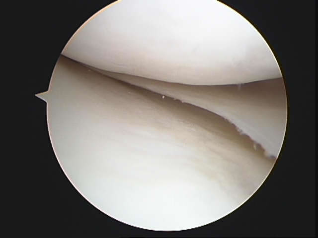 Normal cartilage surfaces
How does it happen?
Cartilage defects may the result of an injury to the knee, but may also occur spontaneously in young children (osteochondritis dissecans)
It may also occur in conjunction with ligament or meniscal injuries.
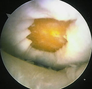 Arthroscopic Picture of a Chondral defect
This can be a potentially serious condition, particularly in young adults, because in the long term it can increase the risk of developing significant osteoarthritis.
What are the symptoms?
The symptoms of chondral injury tend to be pain, clicking, catching and swelling. Quite often the pain is well localized to the area of damage.
Occasionally, it is difficult to say exactly where the pain is arising from, and the knee feels generally uncomfortable.
If the injury has resulted in a fragment of the joint surface cartilage floating around the knee there can also be symptoms of a loose body, with locking and sometimes a palpable lump in the knee that comes and goes.
How is it diagnosed?
The diagnosis of a chondral injury in the knee joint can be made on the story of an injury along with examination of the knee which may reveal nothing more than pain and swelling.
X-rays are useful if there is a small fragment of bone as well as a fragment of articular cartilage (osteochondral lesion). If not, the x-rays will probably be normal.
An MRI scan will show up most chondral injuries.
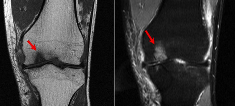 MRI scan of chondral injury
What is the Treatment?
Most acute chondral injuries will be treated with arthroscopic surgery to either remove or replace the damaged fragment.
In general the only repairable injuries of this type are where there is quite a large chunk of articular cartilage with a sliver of bone (an osteochondral fragment) which can be pinned back in place.
If the fragment has to be removed and it leaves behind a crater in an important part of the knee with bare bone in the base, it will not heal up with hyaline cartilage.
It may heal up with fibrocartilage in the base giving some sort of smooth covering.
This type of covering can be encouraged by making small holes in the bone.
By encouraging bleeding into the area, fibrocartilage will form.
This technique is known as microfracture.
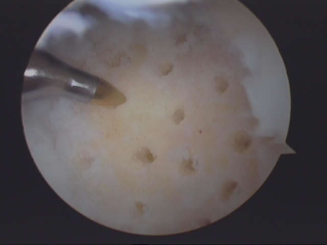 Arthroscopy Picture of Microfracture
If the area of damage is too big to be treated by microfracture, then osteochondral plugs can be taken from other areas of the knee and used to fill the defect - this is a technique known as Mosaicplasty.
Synthetic plugs are also commercially available for Mosaicplasty.
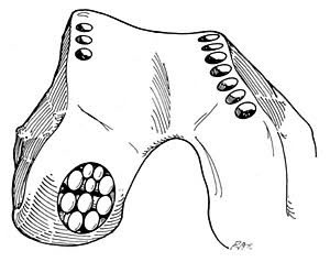 Diagram of Mosaicplasty